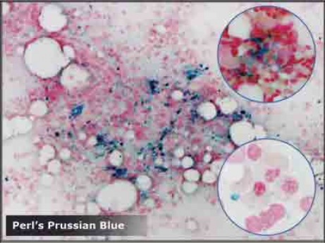
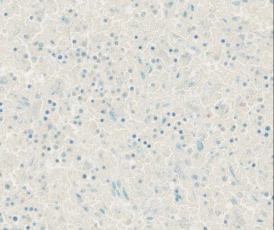
Perl's Prussian Blue
Classification: pigment stain
Mechanism of staining: histochemical
Purpose: stain hemosiderin
Control tissue: spleen, bone marrow
Bone Marrow
|
REAGENT |
PURPOSE |
MECHANISM OF STAINING |
SOURCE OF ERROR |
|
Perl’s solution |
HCl: liberates the ferric ions from the
protein fraction of the hemosiderin molecule Potassium ferrocyanide: combines with ferric ions to produce
ferri-ferrocyanide |
Protein with ferric iron + HCl→ Ferric iron + potassium ferrocyanide
→ Potassium
ferriferrocyanide |
Omitted: Hemosiderin not demonstrated. |
|
Too short: Hemosiderin will be pale. |
|||
|
Too long: Background staining will occur. |
|||
|
Neutral red |
Counterstain: Demonstrates other tissue
components Nuclei & cytoplasm |
Ionic bonding |
Omitted: Collagen, RBC, muscle, cytoplasm
not demonstrated. |
|
Too short: Collagen, RBC, muscle, cytoplasm
too pale. |
|||
|
Too long: Hemosiderin is obscured. |
Special Considerations
Hemosiderin is soluble in acid. Avoid acid fixatives. Also ensure the formalin is buffered and not acidic.
Hemosiderin is an endogenous pigment. When unstained, it will be visible as a yellow or brown pigment.
A positive control is suggested.
Bone Marrow
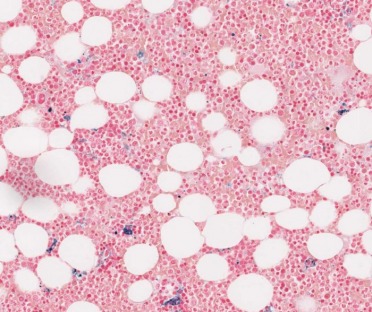
Red: red blood cells, collagen
Dark blue: hemosiderin
Spleen
References
Officer B. HIML251 Lecture notes: Histochemical Stains: Perl’s Prussian Blue & Periodic Acid Schiff, January 14, 2009
Bone Marrow Biopsy- Methodologies. 2008. [cited 2009 April 6].
Available from http://bonemarrowbiopsy.files.wordpress.com/2008/12/perls-prussian-blue.png? w=408&h=306
