Masson Fontana
Classification: pigment stain
Mechanism of staining: histochemical
Purpose: stain hemosiderin
Control tissue: spleen, bone marrow
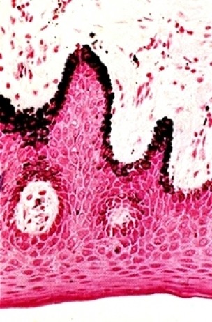
Melanin: black or brown
Nuclei: red
Cytoplasm: light red
Special Considerations:
Argentaffin reactions, unlike argyrophil reactions, require no extraneous reducer.
Rinse glassware before use with distilled water avoid contamination with free metal ions. Also use plastic forceps or wax coated forceps to handle slides.
|
REAGENT |
PURPOSE |
MECHANISM OF STAINING |
SOURCE OF ERROR |
|
Gram’s Iodine |
Oxidation |
Suppresses sulphydryl and tryrosine groups
and exposes phenol groups. |
Omitted: Melanin
will not be demonstrated. |
|
Too short: Trace amounts of melanin may not
be demonstrated. |
|||
|
Too long: Will not affect staining. |
|||
|
5% Sodium thiosulphate |
Bleaching |
Removes brown discoloration |
Omitted: Discoloration in counterstain. Tissue sections will
be discoloured. |
|
Too short: Sections may be slightly
discoloured. |
|||
|
Too long: Will not affect staining. |
|||
|
Methenamine silver (hexamine silver) |
Impregnation o
Silver nitrate o
Hexamine o
Borax |
Forms complexes at the phenol groups |
Omitted: Melanin will not be demonstrated. |
|
Too short: Trace amounts of melanin will not
be detected. |
|||
|
Too long: Non-specific staining may occur. |
|||
|
Gold chloride |
Tones collagen |
Lays down gold in place of silver to remove
any brown colour in other tissue components. Ion exchange |
Omitted: Collagen will remain golden- brown. Toning is
optional. |
|
Too short: Will not affect staining. |
|||
|
Too long: Trace amounts of fungus may not be detected because
gold chloride will start to remove silver staining. |
|||
|
Sodium thiosulphate (hypo) |
Fixation |
Ion exchange of silver with gold. |
Omitted: Non-specific blackening of slide over time due to
remaining unreduced silver. |
|
Too short: Some non-specific blackening of slide over time. |
|||
|
Too long: Will not affect staining. |
|||
|
Nuetral red or Light green |
Counterstain |
Ionic bonding |
Omitted: Background will not be demonstrated. |
|
Too short: Background will be difficult to
see. |
|||
|
Too long: May obscure melanin staining. |
Unbleached Control
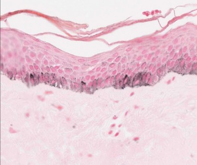
To confirm the presence of melanin, bleaching is performed on a control slide, using a strong oxidizing agent such as potassium permanganate or hydrogen peroxide. This procedure is performed as follows:
· Two consecutive sections are cut
· Bleaching of melanin is done on one slide only
· Perform Masson Fontana technique as per usual on both slides
Bleached Control
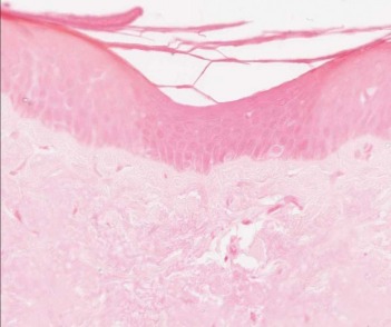
· If the bleached slide is MF negative, and the unbleached slide is MF positive, the tissue likely contains melanin.
References
Officer B. HIML251 Lecture notes: Silver Stains: Grocott’s Methenamine Silver, Jone’s Methenamine Silver, and Masson Fontana, March 18, 2009
University of Vermont Pathology. Masson Fontana Stain for Melanin. 2008. [cited 2009 April 6]. Available from http://education.vetmed.vt.edu/Curriculum/VM8054/Labs/Lab2/Examples/exfontana.htm
Yawney, L. HIML 251 Theory Notes: Masson Fontana. The Michener Institute for Applied Health Sciences, 1989.
