Gomori's Aldehyde Fuschsin
Classification: connective tissue stain
Mechanism of staining: ionic bonding/van der Waals forces
Purpose: stain elastin, beta cells in Islets of Langerhans, basophils in anterior pituitary, and mast cells
Control tissue: aorta, skin, lung, internal control
Properly Stained Slide
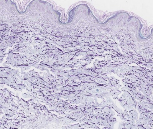
Elastin fibers: Royal purple
Muscle, cytoplasm, collagen: Green
Nuclei: Not demonstrated
|
REAGENT |
PURPOSE |
MECHANISM OF STAINING |
SOURCE OF ERROR |
|
Lugol’s iodine |
Oxidizer |
Creates reactive groups in apolar elastin |
Omitted: Elastin is less intense.
Oxidization is optional. |
|
Too short: Elastin is less intense. |
|||
|
Too long: No problem. |
|||
|
Sodium thiosulphate (hypo) |
Removes iodine discoloration |
|
Omitted: Background discoloration will occur. |
|
Too short: Nuclei and cytoplasm will not be demonstrated or
weakly stained. |
|||
|
Too long: No problem. |
|||
|
Aldehyde fuchsin |
Primary stain (progressive stain) o
Pararosanilin (Basic Fuchsin) o
70% ethanol o
HCl o
Fresh Paraldehyde |
H bonding/ van der Waals bonding to the apolar core of elastin
fibers. Salt linkages to the microbrillar components of elastin. |
Omitted: Elastin not demonstrated. |
|
Too long: : Fine elastin fibers not
demonstrated. |
|||
|
Too short: Fine elastin fibers not
demonstrated. |
|||
|
95% Alcohol rinse |
|
|
Omitted: Aldehyde fuchsin precipitation. |
|
Too long: Fine elastin fibers are not
demonstrated. |
|||
|
Too short: Fine elastin fibers are not demonstrated. |
|||
|
Light green |
Counterstain |
|
Omitted: No other tissue components
demonstrated. |
|
Too long: Fine elastin fibres obscured. |
|||
|
Too short: Other tissues stain lightly and
no detail seen. |
Properly Stained Slide
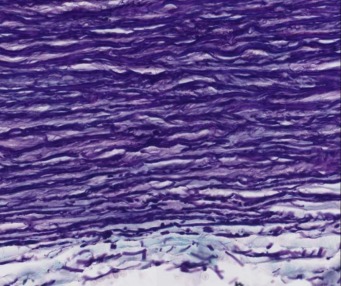
Close up detail of the finely stained royal purple elastin fibers in thin skin.
References
Officer B. HIML251 Lecture notes: Elastic Stains: Verhoeff’s van Gieson & Gomori’s Aldehyde Fuchsin, January 21, 2009
